

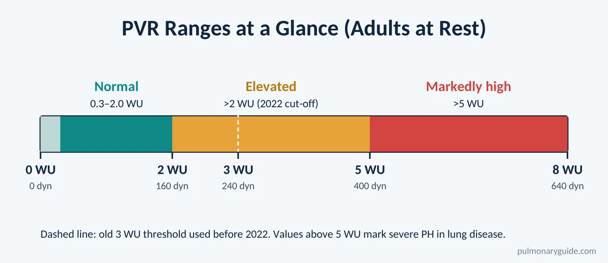
Normal pulmonary vascular resistance in a healthy adult at rest is about 0.3 to 2.0 Wood units (WU), which works out to roughly 24 to 160 dyn·s·cm⁻⁵. Anything above 2 WU is now considered elevated, and it’s one of the key numbers doctors use to confirm and classify pulmonary hypertension.
PVR is calculated with a simple equation: subtract the pulmonary artery wedge pressure from the mean pulmonary artery pressure, then divide by cardiac output. In short, PVR = (mPAP − PAWP) ÷ CO. Multiply the result by 80 if you need it in dyn·s·cm⁻⁵ instead of Wood units.
That’s the quick answer. Below, we’ll walk through what PVR actually measures, how to work out the number step by step, why the “normal” cut-off changed in 2022, and what a high reading means for your lungs and heart.
What Is Pulmonary Vascular Resistance?
Pulmonary vascular resistance is the resistance blood has to push against as it travels from the pulmonary artery, through the tiny vessels of the lungs, and back toward the left side of the heart. Think of it as the “stiffness” or “tightness” of the lung’s blood-vessel network.
Every heartbeat, the right ventricle (the heart chamber that pumps blood into the pulmonary trunk) sends oxygen-poor blood into the lungs. For that to happen easily, the vessels need to be wide, flexible, and relaxed. When they are, resistance stays low and the right ventricle barely has to work. When they narrow, stiffen, or get blocked, resistance rises and the right heart has to pump harder.
The lung circulation is built very differently from the rest of the body. As we explain in our guide to pulmonary and systemic circulation, the lungs handle the entire cardiac output at a fraction of the pressure. According to StatPearls, PVR is only about one-tenth of systemic resistance, which is exactly what allows blood to spread evenly across millions of alveoli for gas exchange.
Several features keep PVR low in healthy lungs:
- Thin-walled vessels with relatively little smooth muscle compared with systemic arteries
- Huge cross-sectional area of the capillary bed
- Spare capacity: closed capillaries can open up (recruitment) and open ones can widen (distension) when blood flow increases
- Constant nitric oxide release from the vessel lining, which keeps the vessels relaxed
If you’d like a refresher on the anatomy, see how the pulmonary arteries branch and carry blood into the lungs.
Normal Pulmonary Vascular Resistance Values
Here are the reference ranges most clinicians use today, based on the 2022 ESC/ERS pulmonary hypertension guidelines.
| Measurement | Normal value | Units |
|---|---|---|
| Pulmonary vascular resistance (PVR) | 0.3–2.0 | Wood units (mmHg·min/L) |
| PVR in metric units | ~24–160 | dyn·s·cm⁻⁵ |
| Total pulmonary resistance (TPR = mPAP ÷ CO) | < 3 | Wood units |
| Mean pulmonary artery pressure (mPAP) | 8–20 | mmHg |
| Pulmonary artery wedge pressure (PAWP) | ≤ 15 | mmHg |
| Cardiac output (CO) | 4–8 | L/min |
Why the Normal Range Changed
For years, a PVR above 3 WU was the threshold for pulmonary hypertension. That’s why many older textbooks still quote a normal range of up to about 250 dyn·s·cm⁻⁵ (roughly 3 WU).
In 2022, the European Society of Cardiology and European Respiratory Society lowered the cut-off. Based on data from healthy people and outcome studies, the guideline states that the upper limit of normal PVR, and the lowest level linked to worse prognosis, is about 2 WU. So a PVR of 2.5 WU, once “borderline normal,” is now considered elevated.
Does Normal PVR Change With Age?
Yes, a little. The ESC/ERS guidelines note that PVR depends on body size and age, and healthy older adults tend to have higher values than younger people. This is one reason doctors interpret a mildly raised number in the context of your age, symptoms, and other test results rather than in isolation.
The Pulmonary Vascular Resistance Formula
The standard pulmonary vascular resistance formula comes from a fluid version of Ohm’s law (resistance = pressure difference ÷ flow):
PVR = (mPAP − PAWP) ÷ CO
Where:
- mPAP = mean pulmonary artery pressure (mmHg), the “inflow” pressure
- PAWP = pulmonary artery wedge pressure (mmHg), a stand-in for left atrial pressure, the “outflow” pressure
- CO = cardiac output (L/min), the total blood flow through the lungs
The top part of the fraction, mPAP − PAWP, is called the transpulmonary pressure gradient (TPG). It tells you how much pressure is “lost” crossing the lung circulation. Dividing it by the flow gives resistance.
You’ll sometimes see the formula for pulmonary vascular resistance written with “PCWP” instead of PAWP. They mean the same thing. Our article on pulmonary capillary wedge pressure explains how that number is obtained and why it reflects left-sided filling pressure.
Converting Wood Units to dyn·s·cm⁻⁵
The result of the equation is in Wood units (mmHg·min/L). To convert to metric resistance units:
PVR (dyn·s·cm⁻⁵) = PVR (WU) × 80
A 2019 review in Clinical Cardiology confirms that one Wood unit equals 80 dyn·s·cm⁻⁵. So 2 WU = 160 dyn·s·cm⁻⁵ and 5 WU = 400 dyn·s·cm⁻⁵.
| Wood units | dyn·s·cm⁻⁵ | Interpretation |
|---|---|---|
| 1.0 | 80 | Normal |
| 2.0 | 160 | Upper limit of normal |
| 3.0 | 240 | Mildly elevated |
| 5.0 | 400 | Significantly elevated |
| 8.0 | 640 | Severely elevated |
Calculating Pulmonary Vascular Resistance: Step by Step
Calculating pulmonary vascular resistance takes only three numbers, all of which come from a right heart catheterization. Here’s how it works in practice.
Step 1: Get the mean pulmonary artery pressure. This is measured directly with a catheter sitting in the pulmonary artery.
Step 2: Get the wedge pressure. The catheter balloon is inflated in a small branch of the pulmonary artery, and the pressure beyond it reflects left atrial pressure.
Step 3: Measure cardiac output. Usually done by thermodilution (averaging at least three readings) or the direct Fick method.
Step 4: Plug the numbers into the equation. Subtract wedge pressure from mean PA pressure, then divide by cardiac output.

Worked Example 1: A Healthy Adult
- mPAP = 15 mmHg
- PAWP = 8 mmHg
- CO = 5 L/min
PVR = (15 − 8) ÷ 5 = 7 ÷ 5 = 1.4 WU (1.4 × 80 = 112 dyn·s·cm⁻⁵)
That sits comfortably within the normal range.
Worked Example 2: Pulmonary Arterial Hypertension
- mPAP = 40 mmHg
- PAWP = 10 mmHg
- CO = 4 L/min
PVR = (40 − 10) ÷ 4 = 30 ÷ 4 = 7.5 WU (600 dyn·s·cm⁻⁵)
This is a markedly raised value with a normal wedge pressure, a pattern typical of pre-capillary pulmonary hypertension such as group 1 pulmonary arterial hypertension.
Worked Example 3: Left Heart Disease
- mPAP = 32 mmHg
- PAWP = 24 mmHg
- CO = 5 L/min
PVR = (32 − 24) ÷ 5 = 8 ÷ 5 = 1.6 WU
Here the pulmonary pressure is high, but the PVR is normal. The problem is back-pressure from the left side of the heart, not disease in the lung vessels themselves. This is called isolated post-capillary PH.
Notice how the same mPAP can mean completely different things depending on PVR. That’s exactly why a pulmonary vascular resistance calculation is required for every patient undergoing a right heart catheter for suspected PH.
Related Calculations: PVRI, TPR and SVR
A few related numbers often appear next to PVR on a catheter report.
Pulmonary Vascular Resistance Index (PVRI)
PVRI adjusts PVR for body size, which matters most in children and in people at the extremes of height and weight. You calculate it with cardiac index instead of cardiac output, which is the same as multiplying PVR by body surface area:
PVRI = (mPAP − PAWP) ÷ CI = PVR × BSA
The correct unit is WU·m², not WU/m². This trips up a lot of people. The Clinical Cardiology review found that more than half of published papers used the wrong units for PVRI, which can lead to real errors in decisions such as whether to close a heart defect. For example, a person with a PVR of 2 WU and a BSA of 2 m² has a PVRI of 4 WU·m², not 1.
Total Pulmonary Resistance (TPR)
TPR = mPAP ÷ CO. It skips the wedge pressure, so it lumps together the resistance of the lung vessels and the back-pressure from the left heart. A normal TPR is under 3 WU.
Systemic Vascular Resistance (SVR) for Comparison
SVR = (mean arterial pressure − right atrial pressure) ÷ CO. In healthy adults it’s usually around 10–20 WU (800–1,600 dyn·s·cm⁻⁵), roughly ten times higher than PVR. This big difference is why we often compare hypertension and pulmonary hypertension as two very different conditions.
| Parameter | Formula | Typical normal |
|---|---|---|
| PVR | (mPAP − PAWP) ÷ CO | 0.3–2.0 WU |
| PVRI | (mPAP − PAWP) ÷ CI | Reported in WU·m² |
| TPR | mPAP ÷ CO | < 3 WU |
| TPG | mPAP − PAWP | Usually < 12 mmHg |
| SVR | (MAP − RAP) ÷ CO | ~10–20 WU |
How Is PVR Measured?
Right Heart Catheterization (Gold Standard)
The only accurate way to calculate pulmonary vascular resistance is with a right heart catheterization. A thin catheter is passed through a vein in the neck, arm, or groin into the right side of the heart and the pulmonary artery. It measures pressures directly and allows cardiac output to be calculated.
The 2022 guidelines recommend that PVR be calculated for every patient having a diagnostic catheter, with pressures recorded at the end of a normal breath out. If you’re curious about the bigger picture, our guides on how pulmonary hypertension is diagnosed and how pulmonary hypertension is measured cover the full work-up.
Echocardiogram Estimates
An echocardiogram can’t measure PVR directly, but cardiologists sometimes estimate it. One widely used method divides the peak tricuspid regurgitation velocity by the velocity-time integral in the right ventricular outflow tract. A ratio above about 0.2 suggests PVR is likely elevated. These estimates are useful for screening and follow-up, but they don’t replace a catheter when a diagnosis or treatment decision depends on the exact number.
What Affects Pulmonary Vascular Resistance?
PVR isn’t a fixed number. It shifts from moment to moment with breathing, posture, oxygen levels, and blood flow.
- Blood flow and pressure. When cardiac output rises, as during exercise, PVR actually falls. Extra capillaries open and existing ones stretch, spreading the flow over a larger area.
- Lung volume. PVR follows a U-shaped curve. It’s lowest at the resting lung volume after a normal breath out (functional residual capacity) and rises at both very low and very high lung volumes.
- Low oxygen. Alveolar hypoxia makes pulmonary arteries constrict. That’s helpful locally, diverting blood to well-ventilated areas, but widespread or chronic hypoxia raises PVR across the whole lung. Read more about hypoxia and pulmonary vasoconstriction.
- Acidosis and high carbon dioxide. Both tend to tighten pulmonary vessels.
- Gravity. In an upright person, resistance is highest at the lung apices and lowest at the bases.
- Chemical signals. Nitric oxide and prostacyclin relax the vessels, while endothelin, thromboxane, and serotonin constrict them. Many PAH medicines target exactly these pathways.
- Blood viscosity. Thicker blood, as in polycythemia, raises resistance.
These factors explain why the lungs normally run at such low pressure. For a broader overview, see our article on the function of pulmonary circulation.
High Pulmonary Vascular Resistance: What It Means
A PVR above 2 WU means the lung vessels are resisting blood flow more than they should. Over time, this forces the right ventricle to work harder, which can lead to right heart enlargement and eventually right heart failure.
PVR and the Definition of Pulmonary Hypertension
Under the 2022 ESC/ERS definitions, pulmonary hypertension is an mPAP above 20 mmHg. PVR and wedge pressure then tell doctors what kind it is:
| Haemodynamic type | mPAP | PAWP | PVR |
|---|---|---|---|
| Pre-capillary PH | > 20 mmHg | ≤ 15 mmHg | > 2 WU |
| Isolated post-capillary PH | > 20 mmHg | > 15 mmHg | ≤ 2 WU |
| Combined post- and pre-capillary PH | > 20 mmHg | > 15 mmHg | > 2 WU |
| Unclassified PH (often high flow) | > 20 mmHg | ≤ 15 mmHg | ≤ 2 WU |
Pre-capillary PH covers PAH, lung-disease-related PH, and chronic thromboembolic PH. Post-capillary PH is the hallmark of group 2 pulmonary hypertension caused by left heart disease. You can see how all five categories fit together in our overview of the groups of pulmonary hypertension.
The guidelines also use higher PVR cut-offs within specific groups. In lung disease, a PVR above 5 WU marks severe PH, which is an important finding in group 3 pulmonary hypertension.
Common Causes of a Raised PVR
- Pulmonary arterial hypertension (idiopathic, heritable, drug-related, or linked to connective tissue disease, HIV, or liver disease)
- COPD, emphysema, and interstitial lung disease
- Chronic low oxygen, including living at high altitude and hypoventilation syndromes
- Chronic blood clots in the lungs (CTEPH)
- Long-standing left heart or mitral valve disease that has started to remodel the lung vessels
- Congenital heart defects with large left-to-right shunts
Why Doctors Care About the Exact Number
PVR guides several major decisions:
- Diagnosis and classification of pulmonary hypertension
- Treatment choice and monitoring. A falling PVR is a good sign that PAH therapy is working. See our guide on how to treat pulmonary arterial hypertension.
- Heart transplant eligibility. A very high, fixed PVR raises the risk that a new heart’s right ventricle will fail. Older transplant criteria treated PVR above 5 WU as a relative contraindication.
- Repairing heart defects. Closing an atrial or ventricular septal defect can be dangerous if PVR is already high.
Persistently high pulmonary artery pressure with a rising PVR is a signal to see a specialist at a pulmonary hypertension centre.
Common Mistakes in the Calculation of Pulmonary Vascular Resistance
Even experienced teams can get this wrong. The most frequent pitfalls in the calculation of pulmonary vascular resistance are:
- Using systolic instead of mean PA pressure. The equation needs the mean.
- Inaccurate wedge pressure. A poorly wedged catheter or reading at the wrong point in the breathing cycle can change the result a lot.
- Unreliable cardiac output. Thermodilution doesn’t work well with intracardiac shunts or severe tricuspid regurgitation, and estimated Fick can be off.
- Mixing up units. Forgetting to multiply by 80, or writing PVRI as WU/m² instead of WU·m².
- Reading the number in isolation. A low PVR with a high mPAP may point to high flow or left heart disease rather than healthy lungs.
Frequently Asked Questions
What is a normal pulmonary vascular resistance?
A normal pulmonary vascular resistance is 0.3 to 2.0 Wood units, or about 24 to 160 dyn·s·cm⁻⁵. Values above 2 WU are considered elevated under current guidelines.
What is the pulmonary vascular resistance equation?
The pulmonary vascular resistance equation is PVR = (mean pulmonary artery pressure − pulmonary artery wedge pressure) ÷ cardiac output. The answer is in Wood units.
How do you convert Wood units to dyn·s·cm⁻⁵?
Multiply by 80. For example, 1.5 WU × 80 = 120 dyn·s·cm⁻⁵. To go the other way, divide by 80.
Is a PVR of 3 Wood units high?
Yes. Since 2022, anything above 2 WU is considered elevated. A PVR of 3 WU is mildly raised and, with an mPAP above 20 mmHg, meets the definition of pre-capillary pulmonary hypertension if wedge pressure is normal.
Can PVR be measured without a heart catheter?
An echocardiogram can estimate whether PVR is likely high, but the only accurate way to calculate it is with a right heart catheterization.
What causes high pulmonary vascular resistance?
Common causes include pulmonary arterial hypertension, chronic lung disease, long-term low oxygen, chronic blood clots in the lungs, and long-standing left heart disease that has remodeled the lung vessels.
Can high PVR be lowered?
Often, yes. Treating the underlying cause, giving oxygen when levels are low, and using PAH medicines such as endothelin receptor antagonists, PDE5 inhibitors, and prostacyclin-pathway drugs can reduce PVR in suitable patients.
What is the difference between PVR and SVR?
PVR is resistance in the lung circulation; SVR is resistance in the rest of the body. SVR is normally about ten times higher than PVR.
The Bottom Line
Normal pulmonary vascular resistance is 0.3–2.0 Wood units (24–160 dyn·s·cm⁻⁵), and a value above 2 WU is now considered abnormal. To calculate it, subtract the wedge pressure from the mean pulmonary artery pressure and divide by cardiac output; multiply by 80 for metric units. On its own, PVR is just a number, but alongside mPAP and wedge pressure it tells doctors whether high lung pressure is coming from the lung vessels, the left heart, or both, which shapes every treatment decision that follows.
Sources
- Widrich J, Shetty M. Physiology, Pulmonary Vascular Resistance. StatPearls, updated 2024.
- Humbert M, Kovacs G, Hoeper MM, et al. 2022 ESC/ERS Guidelines for the diagnosis and treatment of pulmonary hypertension. European Heart Journal, 2022.
- Kwan WC, Shavelle DM, Laughrun DR. Pulmonary vascular resistance index: getting the units right and why it matters. Clinical Cardiology, 2019.
Medical disclaimer: This article is for educational purposes only and isn’t a substitute for professional medical advice. If you have questions about your heart or lung test results, please talk with your doctor or a pulmonary hypertension specialist.

Zachary is a medical assistant who supports the PulmonaryGuide team with research and helps keep our doctor directory accurate and up to date.
More articles by Zachary →

