

Every time your heart beats, it pushes blood on two separate journeys at once. One trip goes out to your whole body. The other is a short, fast loop through your lungs, where blood drops off carbon dioxide and picks up fresh oxygen. That short loop is the answer to what is pulmonary circulation, and it is the reason the air you breathe can reach your brain, muscles and every other organ.
In short: Pulmonary circulation is the flow of blood from the right side of the heart, through the lungs, and back to the left side of the heart. The right ventricle pumps oxygen-poor blood into the pulmonary arteries; in the lung capillaries it releases carbon dioxide and absorbs oxygen; and the pulmonary veins return the oxygen-rich blood to the left atrium, ready to be pumped to the body.
Key Takeaways
- Pulmonary circulation is the heart-to-lungs-to-heart loop. Its main job is gas exchange: oxygen in, carbon dioxide out.
- The pathway runs: right ventricle → pulmonary valve → pulmonary trunk → right and left pulmonary arteries → lung capillaries → pulmonary veins → left atrium.
- It is a low-pressure system. Normal mean pressure in the pulmonary artery is about 14 mmHg, compared with roughly 90 mmHg in the aorta.
- The lungs receive all of the blood the heart pumps, about 5 liters per minute at rest, and can handle several times that during exercise.
- Beyond gas exchange, the lung circulation filters small clots, stores a reserve of blood and helps activate hormones that control blood pressure.
- Pulmonary hypertension, pulmonary embolism and left-sided heart failure are the most common problems that disrupt it.
What Is Pulmonary Circulation? A Simple Definition
Your blood vessels form two circuits that are joined at the heart. The systemic circulation is the big loop: the left ventricle pumps oxygen-rich blood through the aorta to every tissue, and veins bring it back to the right side of the heart. The pulmonary circulation is the small loop: the right ventricle sends that returning, oxygen-poor blood to the lungs to be “recharged,” and it comes back to the left side of the heart to start the big loop again.
“Pulmonary” simply means “relating to the lungs.” Think of the pulmonary circuit as a refueling stop. Blood arrives low on oxygen and loaded with carbon dioxide, spends less than a second next to the air sacs, and leaves topped up. You can see how the two loops connect in our guide to pulmonary and systemic circulation.
The parts involved are:
- Right ventricle – the pump. It is the heart chamber that pumps blood into the pulmonary trunk.
- Pulmonary valve – a one-way door that stops blood from sliding back into the ventricle. Read more about the function of the pulmonary valve.
- Pulmonary trunk – a short, wide vessel (about 5 cm long) that leaves the heart. See the function of the pulmonary trunk.
- Right and left pulmonary arteries – one to each lung, then branching many times. Our article on how many pulmonary arteries there are walks through the branches.
- Pulmonary capillaries – a mesh of tiny vessels wrapped around the alveoli (air sacs), where gas exchange happens.
- Pulmonary veins – usually four, two from each lung, carrying oxygen-rich blood to the heart. Learn about the function of the pulmonary veins.
- Left atrium – the receiving chamber at the end of the loop.
One detail trips up a lot of students: in this circuit, the arteries carry oxygen-poor blood and the veins carry oxygen-rich blood. That sounds backwards, but arteries and veins are named by direction, not oxygen content. Arteries carry blood away from the heart; veins carry it toward the heart.
The Pulmonary Circulation Pathway, Step by Step
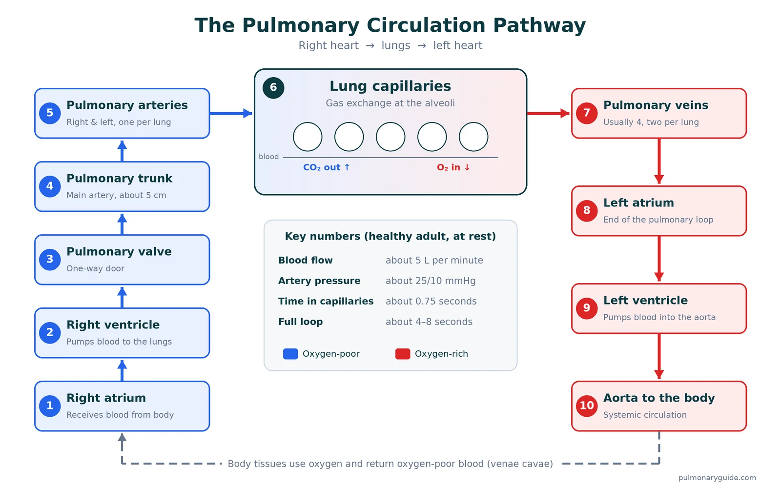
Here is the route a single red blood cell takes, from the moment it returns from the body to the moment it is ready to leave again.
- Return to the right atrium. Oxygen-poor blood from the body enters the right atrium through the superior and inferior vena cava. (Strictly, this is the end of the systemic loop, but it sets up the pulmonary one.)
- Into the right ventricle. Blood passes through the tricuspid valve into the right ventricle.
- Through the pulmonary valve. When the right ventricle contracts, the pulmonary valve opens and blood is pushed into the pulmonary trunk.
- Split to each lung. The trunk divides into the right and left pulmonary arteries. The right one runs a little longer because the heart sits slightly left of center; the left pulmonary artery is shorter.
- Branch, branch, branch. Inside the lungs, the arteries follow the airways and divide into lobar arteries, smaller arteries and arterioles. The pulmonary arteries are thin-walled and stretchy compared with arteries elsewhere.
- Gas exchange in the capillaries. Arterioles feed a dense capillary net around the alveoli. Oxygen diffuses from the air into the blood; carbon dioxide diffuses out. This happens across a barrier far thinner than a sheet of paper. Our page on gas exchange between alveoli and pulmonary capillaries goes deeper.
- Collect into veins. The capillaries drain into venules, which join into larger veins and finally the pulmonary veins.
- Arrive at the left atrium. The pulmonary veins empty into the left atrium. From there, blood flows to the left ventricle and out the aorta to the body.
The whole trip from the right ventricle to the left side of the heart takes only a few seconds, typically about 4 to 8 seconds in a healthy adult at rest. The part that actually matters for breathing, the capillary stretch, takes about three-quarters of a second.
Main Functions of Pulmonary Circulation
Gas exchange gets most of the attention, but the lung circulation does more than swap oxygen and carbon dioxide. Here are its jobs, starting with the big one.
1. Loading oxygen into the blood
Blood arriving in the lungs is only about 75% saturated with oxygen. After one pass through the capillaries, it is close to 100% saturated. Hemoglobin inside red blood cells grabs the oxygen, and the left heart then sends it to the body.
2. Removing carbon dioxide
Carbon dioxide made by your cells travels back to the lungs, mostly as bicarbonate. In the capillaries it is converted back to gas and breathed out. Because carbon dioxide forms acid in the blood, this step also helps keep your blood pH in a narrow, safe range.
3. Filtering the blood
All venous blood passes through the lung capillaries before it reaches the brain or heart. That makes the lungs a natural filter. Tiny clots, small air bubbles and clumps of cells that form in the veins usually get trapped in the lung’s small vessels and broken down, rather than traveling on to cause a stroke. Large clots are a different story: they can block a major pulmonary artery, which is a pulmonary embolism.
4. Acting as a blood reservoir
At any moment, about 500 mL of blood (roughly a tenth of your total) sits in the lung vessels, but only 70 to 100 mL is in the capillaries. Because the vessels are so stretchy, this pool can grow or shrink to smooth out differences between what the right and left ventricles pump, for example when you stand up or take a deep breath.
5. Activating and clearing chemical signals
The inner lining of the lung vessels is coated with angiotensin-converting enzyme (ACE). As blood flows past, ACE turns angiotensin I into angiotensin II, a hormone that raises blood pressure. The same lining also breaks down or removes other substances, such as bradykinin and some serotonin. This is why the lung circulation matters to blood pressure control, not just breathing.
6. Feeding the left side of the heart
The left ventricle can only pump what the lungs deliver. If flow through the lungs drops, say from a large clot or very high lung pressure, the left heart is underfilled and blood pressure falls. This close partnership is covered in our article on the heart and pulmonary artery relationship.
What Makes the Lung Circulation Different
It runs at low pressure
The lungs receive the same amount of blood per minute as the rest of the body combined, yet they do it at about one-fifth of the pressure. Typical pulmonary artery pressure is around 25/10 mmHg, with a mean of about 14 mmHg. Compare that with a normal systemic blood pressure near 120/80 mmHg.
Low pressure is not a weakness; it is the design. The capillary walls must be ultra-thin for gas exchange, and high pressure would push fluid out of them and into the air sacs. That fluid leak is what happens in pulmonary edema.
It has very low resistance
Resistance in the lung vessels is roughly one-tenth of that in the body’s arteries. The vessels have thin walls, little muscle and a huge number of capillaries arranged side by side, so blood meets very little opposition. The capillary bed spreads blood into a sheet about 50 to 100 square meters in area, roughly the floor of a small apartment, and just one red cell thick.
It can open spare capacity on demand
At rest, some lung capillaries carry little or no blood. When flow rises, two things happen: closed capillaries open up (recruitment) and open ones widen (distension). This lets pressure stay low even when blood flow doubles or triples, which is why a healthy person’s lung pressure barely rises during moderate exercise.
It tightens where oxygen is low
In most of the body, low oxygen makes arteries widen. In the lungs it does the opposite. Small pulmonary arteries next to poorly ventilated air sacs constrict, sending blood toward areas that are getting good airflow. This reflex, called hypoxic pulmonary vasoconstriction, keeps air and blood well matched. When oxygen is low throughout the lungs, though, as in severe COPD or at high altitude, the same reflex narrows vessels everywhere and raises lung pressure. Read more in hypoxia and pulmonary vasoconstriction explained.
Gravity matters
Because pressure is so low, gravity has a noticeable effect. When you stand, the bases of your lungs get more blood flow than the tops. Doctors sometimes describe this as lung “zones” based on how artery, vein and air pressure compare at each height.
Pulmonary Circulation by the Numbers
These are approximate values for a healthy adult at rest. Individual results vary, and doctors interpret them alongside symptoms and other tests.
| Measure | Typical value | Why it matters |
|---|---|---|
| Blood flow through the lungs | About 5 L/min (equal to cardiac output) | The lungs are the only organ that receives all of the heart’s output. |
| Pulmonary artery pressure | About 25/10 mmHg | Roughly one-fifth of systemic blood pressure. |
| Mean pulmonary artery pressure | About 14 mmHg | Above 20 mmHg at rest meets the current definition of pulmonary hypertension. |
| Pulmonary capillary wedge pressure | About 4–12 mmHg | Reflects pressure in the left atrium; helps separate heart-related from lung-vessel causes. |
| Blood in the lungs at any moment | About 500 mL | Acts as a reserve that can shift into the body’s circulation. |
| Blood in the capillaries at any moment | About 70–100 mL | Only this thin layer takes part in gas exchange. |
| Capillary transit time | About 0.75 seconds | Oxygen usually equilibrates in about 0.25 seconds, leaving a safety margin. |
| Gas exchange surface | About 50–100 m² | A huge area packed into the chest. |
Want to understand what a high reading means? See high pulmonary artery pressure explained.
Pulmonary vs. Systemic Circulation
| Feature | Pulmonary circulation | Systemic circulation |
|---|---|---|
| Main job | Gas exchange in the lungs | Deliver oxygen and nutrients to tissues |
| Pump | Right ventricle | Left ventricle |
| Starts / ends | Right ventricle → left atrium | Left ventricle → right atrium |
| Arteries carry | Oxygen-poor blood | Oxygen-rich blood |
| Veins carry | Oxygen-rich blood | Oxygen-poor blood |
| Typical pressure | About 25/10 mmHg | About 120/80 mmHg |
| Resistance | Very low | About 10 times higher |
| Response to low oxygen | Vessels constrict | Vessels widen |
| Path length | Short (heart to lungs and back) | Long (whole body) |
Coronary circulation, the blood supply to the heart muscle itself, is a third specialized network. Here is how pulmonary circulation differs from coronary circulation.
Pulmonary vs. Bronchial Circulation: The Lungs Have Two Blood Supplies
This surprises many people: the blood flowing through the pulmonary capillaries is not what keeps lung tissue alive. The airways, lung connective tissue and the walls of large vessels get their own oxygen-rich blood from the bronchial arteries, small branches of the aorta.
- Pulmonary circulation: high flow, low pressure, comes from the right heart, exists for gas exchange.
- Bronchial circulation: low flow (about 1–2% of cardiac output), systemic pressure, comes from the aorta, exists to nourish lung tissue.
This double supply helps explain why a pulmonary embolism does not always kill the lung tissue beyond the clot. The bronchial arteries can keep it partly supplied.
How Pulmonary Circulation Changes Before and After Birth
Before birth, a baby gets oxygen from the placenta, not the lungs. The lungs are filled with fluid, the pulmonary vessels are tightly constricted and resistance is high. Two shortcuts let most blood skip the lungs:
- Foramen ovale – an opening between the right and left atria.
- Ductus arteriosus – a vessel linking the pulmonary trunk to the aorta.
With the first breaths, the lungs fill with air, oxygen levels rise and the lung vessels relax. Pulmonary resistance drops sharply, blood rushes into the lungs, and both shortcuts close over the following hours to weeks. If this switch fails, a newborn can develop persistent pulmonary hypertension, a condition that needs urgent care in a neonatal unit.
Pulmonary Circulation During Exercise
During hard exercise, cardiac output in a fit adult can rise to four or more times its resting level, and all of that extra blood must pass through the lungs. Recruitment and distension of capillaries let the lungs take this load with only a modest rise in pressure. Blood moves faster, so time in the capillaries can drop toward a third of a second, which is still enough for healthy lungs to load oxygen fully.
In people with lung disease or stiff, narrowed pulmonary vessels, that safety margin disappears. Oxygen levels may be normal at rest but fall during walking or climbing stairs. This is one reason doctors use exercise tests such as a six-minute walk.
Conditions That Disrupt Pulmonary Circulation
Problems in the lung circulation usually show up in one of three ways: pressure goes up, flow is blocked, or gas exchange fails.
| Condition | What goes wrong | Common signs |
|---|---|---|
| Pulmonary hypertension | Pressure in the lung arteries stays too high, straining the right ventricle | Breathlessness on exertion, fatigue, chest pain, ankle swelling, fainting |
| Pulmonary embolism | A clot, usually from a leg vein, blocks a pulmonary artery | Sudden shortness of breath, sharp chest pain, fast heartbeat, coughing blood |
| Left-sided heart failure or mitral valve disease | Blood backs up into the pulmonary veins and capillaries | Breathlessness lying flat, waking up short of breath, crackles in the lungs |
| Pulmonary edema | Fluid leaks from capillaries into the air sacs | Severe breathlessness, frothy or pink sputum |
| Chronic lung disease (COPD, fibrosis) | Low oxygen narrows vessels; scarring destroys capillaries | Long-term breathlessness, low oxygen levels |
| Congenital heart defects | Holes or abnormal connections send too much blood to the lungs | Poor growth in children, breathlessness, bluish skin in advanced cases |
Pulmonary hypertension is grouped into five types by cause; in the United States, the most common cause is left-sided heart disease (NHLBI). Learn more about the groups of pulmonary hypertension and the causes of pulmonary hypertension. For clots, see how to know if you have a pulmonary embolism.
How Doctors Check Your Pulmonary Circulation
No single test covers everything. Doctors usually combine several:
- Pulse oximetry and arterial blood gas – show how well blood is being oxygenated.
- Echocardiogram – an ultrasound of the heart that estimates pulmonary artery pressure and checks the right ventricle. Often the first test for suspected pulmonary hypertension.
- CT pulmonary angiogram – a contrast CT scan that shows clots in the pulmonary arteries.
- V/Q scan – compares airflow and blood flow in each part of the lungs; useful for chronic clots.
- Pulmonary function tests with DLCO – measure how easily gas moves from the air sacs into the blood.
- Right heart catheterization – a thin tube threaded into the pulmonary artery to measure pressures directly. This is the test that confirms pulmonary hypertension.
For a walkthrough of the testing process, see how pulmonary hypertension is diagnosed.
Keeping Your Pulmonary Circulation Healthy
- Don’t smoke or vape. Smoking damages lung capillaries and airways and raises clot risk.
- Move regularly. Aim for about 150 minutes of moderate activity a week, unless your doctor advises otherwise.
- Break up long periods of sitting. On flights and long drives, get up or flex your calves often to lower the risk of leg clots.
- Treat sleep apnea. Repeated drops in oxygen overnight can raise lung pressure over time.
- Control blood pressure and heart disease. A healthy left heart keeps blood from backing up into the lungs.
- Take symptoms seriously. Breathlessness that is new or getting worse deserves a checkup, not just more rest.
When to See a Doctor
Make an appointment with your doctor or a lung specialist if you notice:
- Shortness of breath with everyday activity that is new or worsening
- Tiredness that doesn’t improve with rest
- Swelling in your ankles, legs or belly
- Lightheadedness or near-fainting when you exert yourself
- A bluish tint to your lips or fingertips
You can search for a pulmonologist near you in our doctor directory.
Call 911 or go to the nearest emergency room right away if you have sudden shortness of breath, chest pain that gets worse when you breathe in, coughing up blood, fainting, or a racing heartbeat, especially after surgery, a long trip or a period of bed rest. These can be signs of a pulmonary embolism, which is a medical emergency.
Frequently Asked Questions
What is pulmonary circulation in simple terms?
It is the loop that carries blood from your heart to your lungs and back. The right side of the heart sends used, oxygen-poor blood to the lungs, the blood picks up oxygen and drops off carbon dioxide, and it returns to the left side of the heart to be pumped to the body.
What is the main function of pulmonary circulation?
Its main function is gas exchange: adding oxygen to the blood and removing carbon dioxide. It also filters small clots, holds a reserve of blood and helps activate the hormone angiotensin II.
Where does pulmonary circulation start and end?
It starts in the right ventricle, which pumps blood through the pulmonary valve into the pulmonary trunk. It ends in the left atrium, where the pulmonary veins deliver oxygen-rich blood.
Why do pulmonary arteries carry deoxygenated blood?
Arteries are defined by direction, not oxygen. Any vessel carrying blood away from the heart is an artery. The pulmonary arteries carry blood away from the heart toward the lungs, and that blood hasn’t picked up oxygen yet.
Why is pulmonary circulation low pressure?
The lung capillaries must be extremely thin to let gases pass, and the lungs only need to push blood a short distance. Low pressure keeps fluid from leaking into the air sacs while still moving the full cardiac output.
How long does it take blood to go through the lungs?
The trip from the right ventricle to the left side of the heart takes roughly 4 to 8 seconds at rest. Each red blood cell spends only about 0.75 seconds in the capillaries where gas exchange happens.
What is the difference between pulmonary and bronchial circulation?
Pulmonary circulation comes from the right heart and exists for gas exchange. Bronchial circulation comes from the aorta and supplies oxygen and nutrients to the lung tissue itself, such as the airways.
The Bottom Line
If you have ever wondered what is pulmonary circulation, the short answer is that it is your body’s oxygen refueling loop: right heart, lungs, left heart, in a few seconds, with every heartbeat. It works at low pressure, adjusts automatically to exercise and oxygen levels, and quietly filters and conditions your blood along the way. When it falters, the first sign is usually breathlessness, so new or worsening shortness of breath is always worth checking. To see how this loop fits into breathing as a whole, read how the pulmonary system works.
References
- Physiology, Pulmonary Circulatory System. StatPearls, NCBI Bookshelf. https://www.ncbi.nlm.nih.gov/books/NBK525948/
- Circulatory System. Cleveland Clinic. https://my.clevelandclinic.org/health/body/circulatory-and-cardiovascular-system
- Pulmonary Arteries. Cleveland Clinic. https://my.clevelandclinic.org/health/body/21486-pulmonary-arteries
- What Is Pulmonary Hypertension? National Heart, Lung, and Blood Institute (NHLBI). https://www.nhlbi.nih.gov/health/pulmonary-hypertension
- Circulatory Pathways. SEER Training Modules, National Cancer Institute. https://training.seer.cancer.gov/anatomy/cardiovascular/blood/pathways.html
- Humbert M, et al. 2022 ESC/ERS Guidelines for the diagnosis and treatment of pulmonary hypertension. European Heart Journal. 2022;43(38):3618–3731.
This article is for general education and is not a substitute for advice from your doctor.

Dr. Dennis Rodman is a physician on the PulmonaryGuide medical team. He medically reviews our articles for accuracy before they are published and writes about COPD, pulmonary embolism and children's lung health.
More articles by Dennis →

