

The function of a pulmonary vein is to carry freshly oxygenated blood from the lungs back to the heart. Pulmonary veins are the last link in the lung circuit: they collect blood that has just picked up oxygen in the air sacs and deliver it to the left atrium, which passes it to the left ventricle to be pumped out to the whole body.
That makes them unusual. They are the only veins in an adult that carry oxygen-rich blood, and most people have four of them. Below, you’ll see exactly where they carry blood, how they’re built, how they differ from pulmonary arteries, and why heart rhythm specialists pay so much attention to them.
Key Takeaways
- Pulmonary veins carry oxygen-rich blood from the lungs to the left atrium of the heart.
- Most people (about 60–70%) have four: right superior, right inferior, left superior and left inferior. Three or five is a normal variation.
- They are called veins because they carry blood toward the heart, not because of the blood’s oxygen level.
- Unlike most leg and arm veins, pulmonary veins have no valves. Pressure in them closely tracks pressure in the left atrium.
- Heart muscle extends a short way into each vein. This “sleeve” is where more than 90% of atrial fibrillation triggers start, which is why AFib ablation targets the pulmonary veins.
- Problems include back-pressure from left heart disease, narrowing (stenosis), clots and birth defects such as TAPVR.
What Is the Function of a Pulmonary Vein?
Think of your circulation as two loops joined at the heart. The pulmonary and systemic circulations run one after the other, and the pulmonary veins are the hand-off point between them.
The right side of the heart pumps oxygen-poor blood out through the pulmonary arteries into the lungs. In the lungs, that blood passes through capillaries wrapped around millions of tiny air sacs (alveoli), where it releases carbon dioxide and picks up oxygen. The pulmonary veins then gather this refreshed blood and bring it back to the left side of the heart so it can be sent out to the brain, muscles and every other organ.
In practical terms, the function of a pulmonary vein comes down to three jobs:
- Return oxygenated blood. Every drop of blood your left ventricle pumps to the body has just come through the pulmonary veins.
- Act as a low-pressure collecting system. Thousands of small lung venules merge into larger and larger branches, ending in the main veins that enter the heart.
- Connect the lungs to the left heart without valves. Because there’s no valve in the way, pressure changes in the left atrium pass straight back into the lungs. That fact explains a lot of lung symptoms in heart failure (more on this below).
Where Do the Pulmonary Veins Carry Blood? Step by Step
Here is the full path a red blood cell takes through the lung circuit, with the pulmonary veins’ part highlighted:
- Right atrium: Oxygen-poor blood returns from the body through the superior and inferior vena cava.
- Right ventricle: It pumps blood through the pulmonary valve into the pulmonary trunk. (See which heart chamber pumps blood into the pulmonary trunk.)
- Pulmonary arteries: The trunk splits into right and left pulmonary arteries, one for each lung.
- Lung capillaries: Gas exchange between the alveoli and capillaries loads oxygen and unloads carbon dioxide. This takes less than a second.
- Pulmonary veins: Small venules join into larger veins that leave each lung at its root (the hilum), usually as two veins per lung.
- Left atrium: The four veins empty into the back wall of the left atrium through four separate openings (ostia).
- Left ventricle and aorta: Blood passes through the mitral valve into the left ventricle, which pumps it into the aorta and out to the body.
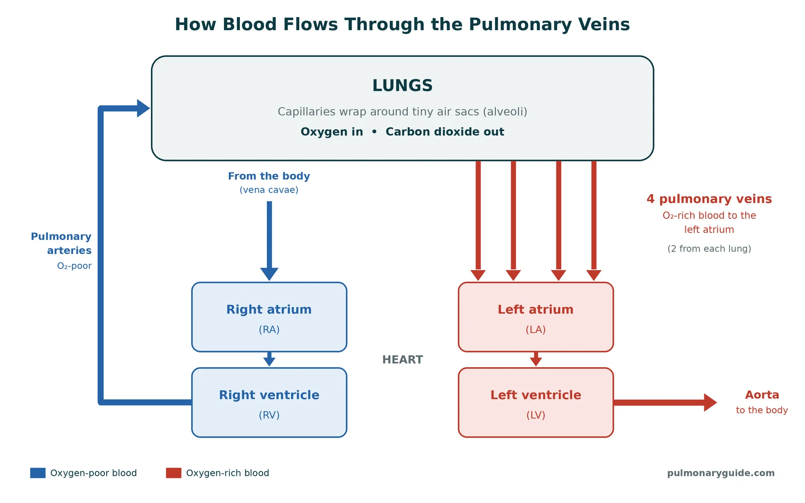
A useful way to remember it: arteries go away from the heart, veins go toward it. Oxygen level has nothing to do with the name.
Pulmonary Vein Anatomy: The Four Main Veins
Most people have two pulmonary veins from each lung, named for their side and position (superior = upper, inferior = lower). Our guide to how many pulmonary veins there are covers the numbers in more detail.
| Vein | Drains | Notes |
|---|---|---|
| Right superior pulmonary vein | Right upper lobe and right middle lobe | The middle lobe sometimes has its own separate vein |
| Right inferior pulmonary vein | Right lower lobe | Usually the lowest structure at the right lung root |
| Left superior pulmonary vein | Left upper lobe and lingula | Most often the source of AFib triggers |
| Left inferior pulmonary vein | Left lower lobe | May join the left superior vein in a common trunk |
Size and structure
A healthy main pulmonary vein measures roughly 9 to 13 mm across, about the width of a pencil to a small fingertip. Like other veins, the wall has three layers: a smooth inner lining (endothelium), a middle layer of smooth muscle, and a tough outer layer of connective tissue.
Two features set them apart:
- No valves. Veins in your legs have one-way valves to fight gravity. Pulmonary veins don’t need them, so blood and pressure can move freely between the lungs and the left atrium.
- A sleeve of heart muscle. Atrial heart muscle extends into the first part of each vein, on average about 9 mm and sometimes a few centimeters. Superior veins tend to have longer sleeves. This is the tissue involved in atrial fibrillation.
Normal variations
Anatomy textbooks show four tidy veins, but real people vary. According to StatPearls (NIH), the classic four-vein pattern appears in about 60–70% of people. The rest have harmless variants, most commonly:
- A left common trunk: the two left veins merge before entering the heart, giving one opening instead of two. A short common trunk is seen in roughly 15% of people.
- An extra right vein: often a separate middle lobe vein, giving three veins on the right.
These variants don’t cause symptoms. They matter mainly to surgeons and to electrophysiologists planning an ablation, who map the veins with CT or MRI beforehand.
Pulmonary Veins vs. Pulmonary Arteries vs. Other Veins
The lung circuit flips the usual rules about oxygen, which confuses a lot of students. This table lines them up side by side:
| Feature | Pulmonary veins | Pulmonary arteries | Most other (systemic) veins |
|---|---|---|---|
| Direction of flow | Lungs → heart | Heart → lungs | Body → heart |
| Oxygen content | High (about 95–100% saturated) | Low (about 70–75% saturated) | Low |
| Heart chamber connected | Left atrium | Right ventricle | Right atrium (via the vena cavae) |
| Typical pressure | About 6–12 mmHg (mirrors left atrial pressure) | Mean 20 mmHg or less | About 2–6 mmHg near the heart |
| Valves | None | Pulmonary valve at the start | Many, especially in the legs |
| Number of main vessels | Usually 4 | 1 trunk splitting into 2 | Many |
Why isn’t pulmonary vein blood a perfect 100%? A small amount of oxygen-poor blood from the bronchial veins, which drain the airway walls, empties into the pulmonary veins. This “physiologic shunt” nudges the saturation of blood reaching the left atrium just below 100%.
For a wider view of how the lung circuit compares with the heart’s own blood supply, see how pulmonary circulation differs from coronary circulation.
More Than a Pipe: Lesser-Known Roles of the Pulmonary Veins
Most articles stop at “they carry oxygenated blood.” In clinical practice, the pulmonary veins also matter in three less obvious ways.
1. A window into left heart pressure
Because there’s no valve between the pulmonary veins and the left atrium, the pressure in the veins rises and falls with left atrial pressure. Doctors use this during right heart catheterization: by wedging a balloon catheter in a small lung artery, they record a pressure that reflects the pulmonary veins and left atrium. That number is the pulmonary capillary wedge pressure (PCWP), normally about 6–12 mmHg.
2. The source of lung congestion in heart failure
When the left side of the heart can’t keep up, as in left heart failure or mitral valve disease, pressure backs up into the pulmonary veins and then the lung capillaries. Fluid leaks into lung tissue, causing breathlessness, especially when lying flat. This is called pulmonary venous hypertension, and when it raises lung artery pressures too, it’s classed as Group 2 pulmonary hypertension, the most common of the five groups of pulmonary hypertension.
3. An electrical trigger zone
The muscle sleeves at the vein openings can fire off stray electrical signals. In some people those signals set off atrial fibrillation, which is why the pulmonary veins are the main target of AFib ablation (next section).
The veins also have a dense nerve supply near the heart and smooth muscle in their walls, so they can change tone slightly. Their role here is far smaller than the arteries’, which tighten when oxygen is low through hypoxic pulmonary vasoconstriction.
Pulmonary Veins and Atrial Fibrillation
Atrial fibrillation (AFib) is the most common sustained heart rhythm problem, affecting more than 2 million adults in the United States. In the late 1990s, researchers in France discovered that the extra beats that start AFib come from the pulmonary veins in more than 90% of cases, usually from the muscle sleeves near the vein openings.
That finding led to pulmonary vein isolation (PVI), now the cornerstone of AFib ablation. Here’s how it works in plain terms:
- A cardiologist threads thin catheters from a vein in the groin up into the left atrium.
- Using heat (radiofrequency) or extreme cold (cryoablation), they create a ring of scar tissue around the opening of each pulmonary vein.
- Scar doesn’t conduct electricity, so stray signals from inside the veins can no longer reach the rest of the heart.
PVI is generally safe. Cleveland Clinic lists a roughly 1% risk of pulmonary vein damage, which can lead to narrowing of the vein (pulmonary vein stenosis). New breathlessness, cough or chest pain in the weeks or months after ablation should be reported to your care team.
Conditions That Affect the Pulmonary Veins
Pulmonary vein problems are uncommon compared with artery problems like pulmonary embolism, but they can be serious.
| Condition | What happens | Who is usually affected |
|---|---|---|
| Pulmonary venous hypertension | Pressure backs up from the left heart into the veins and lungs | Adults with left heart failure or mitral valve disease |
| Pulmonary vein stenosis | One or more veins narrow, slowing blood return and raising lung pressure | Infants (often with other heart defects); adults after AFib ablation or from outside compression |
| Pulmonary vein thrombosis | A clot forms inside a pulmonary vein | Rare; mainly after lung surgery or transplant, after ablation, or with lung cancer |
| Total anomalous pulmonary venous return (TAPVR) | All pulmonary veins connect to the right side of the heart instead of the left | Newborns; needs surgery |
| Partial anomalous pulmonary venous return (PAPVR) | One or more (but not all) veins connect to the wrong place | Children or adults; may go unnoticed for years |
| Tumor compression or invasion | A lung or heart tumor blocks a vein | Adults with lung or cardiac tumors |
In TAPVR, oxygen-rich blood ends up mixing with oxygen-poor blood on the right side of the heart, so babies may look bluish (cyanosis), breathe fast and feed poorly. A hole between the upper heart chambers usually keeps them alive until surgeons reconnect the veins to the left atrium. Untreated, about 80% of babies with TAPVR die in the first year, so it is repaired early, and most children do well afterward.
A common point of confusion: a pulmonary embolism is not a pulmonary vein problem. A PE is a clot that travels from the legs through the right heart and lodges in the pulmonary arteries. If you’re worried about that, read how to tell if you have a pulmonary embolism.
How Doctors Check the Pulmonary Veins
No single blood test checks pulmonary vein function. Doctors rely on imaging and pressure measurements:
- Echocardiogram: an ultrasound of the heart. It shows blood flow from the veins into the left atrium and is the first test for suspected TAPVR in babies.
- CT angiography (CT venography): gives the most detailed 3D map of the veins and is standard before AFib ablation.
- Cardiac MRI: maps vein anatomy and blood flow without radiation.
- Chest X-ray: can show lung congestion or the curved “scimitar” vein seen in one form of PAPVR.
- Right heart catheterization: measures wedge pressure and lung artery pressure. It’s also how doctors diagnose pulmonary hypertension.
When to See a Doctor
You can’t feel your pulmonary veins working, and most people never have a problem with them. Book an appointment with your doctor if you notice:
- Shortness of breath that is new, getting worse, or happens with light activity
- Breathlessness when lying flat, or waking up at night short of breath
- A racing, fluttering or irregular heartbeat
- Swelling in the ankles or legs along with breathlessness
- New cough, breathlessness or chest discomfort in the weeks or months after an AFib ablation
- In a baby: fast breathing, bluish lips or skin, poor feeding or poor weight gain
Call 911 right away for sudden severe shortness of breath, chest pain or pressure, coughing up blood, fainting, or blue or gray lips. These can signal a medical emergency.
If you need a lung specialist, you can find a pulmonologist near you in our directory. Heart rhythm problems are usually managed by a cardiologist or electrophysiologist.
Frequently Asked Questions
What is the main function of a pulmonary vein?
The main function of a pulmonary vein is to carry oxygen-rich blood from the lungs to the left atrium of the heart. From there, the left ventricle pumps it to the rest of the body.
Why is it called a vein if it carries oxygenated blood?
Blood vessels are named by direction, not oxygen content. Veins carry blood toward the heart and arteries carry it away. Pulmonary veins carry blood toward the heart, so they’re veins, even though their blood is oxygen-rich.
Where do the pulmonary veins carry blood to?
They carry blood to the left atrium, the upper-left chamber of the heart. Normally each vein enters through its own opening in the back wall of the left atrium.
How many pulmonary veins do humans have?
Most people have four: two from each lung. About a third of people have a normal variation with three or five, such as two left veins joining into one trunk or an extra right vein. These variations usually cause no problems.
What is the difference between a pulmonary vein and a pulmonary artery?
Pulmonary arteries carry oxygen-poor blood from the right ventricle to the lungs. Pulmonary veins carry oxygen-rich blood from the lungs to the left atrium. They are the reverse of the body’s other arteries and veins. Learn more in our guide to the function of pulmonary circulation.
Do pulmonary veins have valves?
No. Pulmonary veins have no valves where they join the left atrium. That’s why high pressure in the left heart passes straight back into the lungs and can cause fluid buildup.
What happens if a pulmonary vein is blocked or narrowed?
Blood from that part of the lung can’t drain properly, so pressure rises in the lung vessels. People may develop shortness of breath, cough, reduced exercise ability, coughing up blood or pulmonary hypertension. Treatment depends on the cause and can include balloon widening, stents or surgery.
The Bottom Line
Pulmonary veins are short, valveless vessels with a big job: they bring every bit of freshly oxygenated blood from your lungs into the left side of your heart. Beyond that core function, they act as a pressure link between the left heart and the lungs and as the starting point for most atrial fibrillation. If you’d like to see how they fit with the rest of the system, our overview of how the pulmonary system works and the function of the pulmonary circuit are good next reads.
References
- Poschel D, Bordoni B. Anatomy, Thorax, Lung Veins. StatPearls. Updated July 26, 2025.
- Cleveland Clinic. Pulmonary Veins: Anatomy and Function.
- Cleveland Clinic. Pulmonary Vein Isolation (PVI) Ablation.
- National Heart, Lung, and Blood Institute. What Is Atrial Fibrillation?
- Mayo Clinic. Total anomalous pulmonary venous return (TAPVR).
- Haïssaguerre M, et al. Spontaneous initiation of atrial fibrillation by ectopic beats originating in the pulmonary veins. N Engl J Med. 1998;339(10):659-666.
- Bunch TJ, Cutler MJ. Is pulmonary vein isolation still the cornerstone in atrial fibrillation ablation? J Thorac Dis. 2015;7(2):132-141.
This article is for general education and is not a substitute for advice from your doctor.

Dr. Salman Rayhan is a physician on the PulmonaryGuide medical team. He medically reviews our articles for accuracy before they are published and writes about pulmonary embolism diagnosis and treatment and lung cancer care.
More articles by Salman →

